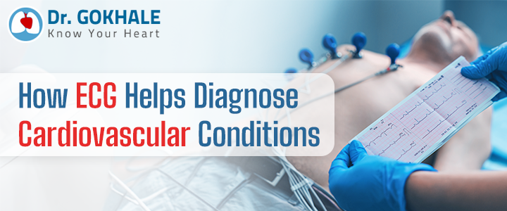
Electrocardiogram, simply ECG, is one of the most commonly heard medical terms, especially regarding heart-related ailments or diagnoses.
One of the most instrumental diagnostic technologies, ECG can identify minor irregularities and detect serious cardiac issues. It has helped cardiologists’ worldwide gain key information about the heart’s function, something impossible before its invention.
ECG for Heart Disease Detection: A Cornerstone in Modern Cardiology:
Relying on symptoms was the only way to evaluate the activity of the heart’s electrical function until the invention of ECG which provided an accurate, painless, and non-invasive alternative.
ECG is quite possibly a landmark invention that became a cornerstone of cardiology and paved the way for more advanced cardiac diagnostics and treatment options. While it is a significant invention and a commonly heard key diagnostic tool, not many people truly know how it works or what heart-related condition it helps diagnose.
In this blog, we explore these questions with insights from Dr. Alla Gopala Krishna Gokhale, a leading heart transplant surgeon in Hyderabad. Today, we offer a brief yet critical look into how ECG works and what types of heart-related conditions it can help diagnose.
Electrocardiogram for Heart Health – How Does an ECG Work?
Our heartbeat begins with an electrical impulse from the heart’s natural pacemaker, the sinoatrial (SA) node. This impulse triggers the heart muscles to contract and relax in a rhythmic cycle, allowing it to pump blood throughout the body.
This electrical activity moves through the heart in wave-like patterns that can be detected on the surface of the skin. An ECG (electrocardiogram) captures these waves, thereby helping us to measure and monitor the heart’s electrical activity.
Any change in heart health can affect this electrical activity. And these changes are often reflected in the ECG, thus helping heart specialists detect irregularities early to ensure timely diagnosis and care.
ECG for Heart Health: How ECG Helps Diagnose Cardiovascular Conditions?
During an ECG test, electrodes (12) are attached to specific areas of the chest, arms, and legs using a small sticky patch wired to the instruments. As the heart beats, the machine visually represents electrical impulses through graphs with waves and intervals.
The shape, size, and changes in the waves concerning timing and rhythm give away the abnormalities in the heart, which are easily spotted. Here are the different types of heart-related conditions that are identified using ECG as a diagnostic tool:
Arrhythmias (Irregular Heart Rhythms):
Arrhythmia is a condition in which the heart beats too fast, too slow, or unpredictably. ECG is the frontline tool for identifying this abnormality, as changes in the heartbeat are easily recognized in the electrical activity.
Heart rate changes, irregular heartbeats (like premature beats), conduction problems, and disruptions such as delays or blocks – each type of arrhythmia has a signature ECG pattern easily recognized and diagnosed by cardiologists in Hyderabad.
Heart Attacks (Myocardial Infarctions):
As the heart’s electrical activity varies during and after an attack, ECG is also a primary tool used to detect and provide vital insights about these incidents.
For example, in cases of active heart damage, this change is reflected in changes in the ST segments and T waves of the ECG due to restricted blood flow.
When a heart region is affected, the electrical activity does vary. As the electrodes are placed at different locations on the chest, the extent of the damage to the specific region can also be gauged using the ECG reading recorded.
Even in the case of previous silent heart attacks, the presence of deep Q waves in ECG waves recorded by particular leads may indicate past damage, even if the person never experienced obvious symptoms.
Coronary Artery Disease (Blocked Arteries):
Although someone with stable coronary artery disease might show a normal ECG at rest, specific changes can appear when symptoms strike or during a stress test. Indicators include:
- ST segment depression, often seen when the heart muscle doesn’t receive enough oxygen.
- Inverted T waves, which may also suggest temporary or chronic ischemia.
- Abnormal Q waves, pointing to old infarctions caused by previously blocked arteries.
These subtle alterations detected by the heart specialist often give early warnings before more serious events occur.
Heart Muscle Thickening (Hypertrophy):
Conditions like chronic high blood pressure or inherited heart diseases can cause the heart muscle to thicken, and these changes can also be easily identified on the ECG.
The changes often show up as:
- Increased QRS voltage, as a bulkier muscle generates stronger electrical signals.
- ST and T wave abnormalities reflect changes in how the heart resets between beats.
- Widened or unusual P waves, potentially indicating enlargement of the left atrium.
These patterns help cardiologists identify structural changes even before symptoms develop.
Electrolyte Imbalances:
“Electrolytes such as potassium, calcium, and magnesium are essential to heart rhythm stability. Their imbalances often lead to noticeable, unique shifts on the ECG, which the cardiologists easily detect”, says heart transplant doctor in Hyderabad Dr Gopala Krishna Gokhale.
Pericarditis (Heart Lining Inflammation):
When the pericardium, the membrane surrounding the heart, becomes inflamed, it produces distinct ECG changes due to subtle changes in conduction. These subtle changes in the ECG include:
- ST-segment elevation across multiple leads contrasts with the localized elevations seen in heart attacks.
- Depressed PR segments, a classic marker of pericarditis.
- Inverted T waves, typically appearing later in the condition’s course.
When paired with symptoms like chest pain and fever, these signs help heart specialists detect and distinguish pericarditis from more dangerous causes of chest discomfort.
In addition to the above, the ECG (electrocardiogram) is also an essential tool for heart transplant surgeons to monitor heart health before, during, and after the surgery.
“ECG helps establish a baseline of the recipient’s heart rhythm and identify any pre-existing arrhythmias before heart transplant surgery. It provides continuous monitoring of the electrical activity of both the recipients and donor’s hearts during, and monitor the rhythm of the transplanted heart and also detect signs of rejection”, concludes the best heart transplant surgeon in Hyderabad, Dr Alla Gopala Krishna Gokhale.
 Ask Doctor
